The HRT: Examining the Optic Nerve in GlaucomaGlaucoma specialists can get a good idea of the amount of
cupping in an optic nerve by looking at it with an instrument known as an
ophthalmoscope. They can get an idea of whether the cupping is remaining
stable or worsening by taking a series of photographs over time. But these
methods have an important limitation. They can only suggest how big the
cup is in the same way that an ordinary aerial photograph of a crater
could can give us only a rough idea of how deep the crater is. We can get
a much better idea of the depth of the cup or crater by taking a
stereoscopic photograph. This would allow us actually to measure just how
much the optic nerve has been damaged.
The HRT and How It Works
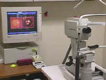
Glaucoma Service doctors are now examining patients with
an instrument that can give more detailed information about the
3-dimensional structure of the cup -- the Heidelberg Retina Tomograph
(HRT). The HRT uses a special laser to take 3-dimensional photographs of
the optic nerve and surrounding retina.
This laser, which is not powerful enough to harm the eye,
is first focused on the surface of the optic nerve and captures that
image. Then it is focused on the layer just below the surface and captures
that image. The HRT continues to take images of deeper and deeper layers
until the desired depth has been reached. Finally, the instrument takes
all these pictures of the layers and puts them together to form a
3-dimentional image of the entire optic nerve.
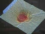
The HRT takes 32 layer-by-layer pictures from the surface
of the optic nerve to from 0.5 mm to 4.0 mm deep into the ocular
structures. The computer then piles all the slices together in a
reconstructed paper printout that looks like a map drawn to represent the
hills and valleys of a geographical area. By color coding areas of
elevation and depression, the HRT provides a two-dimensional
representation of what the original, three-dimensional, stack looks like.
The HRT image can be used to compute things such as the
area of the optic disc (the part of the optic nerve at the back of the
eye), the volume of the cup, and the area of the rim around the cup as
well. These numbers can then be used in two ways. First, they might show
measurements different enough from normal to help in diagnosing glaucoma.
As changes in the optic nerve are often the first sign of glaucoma and can
precede visual field changes, one might be able to diagnose the disease
earlier. Second, the measurements can be followed over time by taking a
series of tests - much like taking a series of visual fields. Changes in
depth are then computed.Various changes might indicate a worsening or
mprovement in the disease.
|
 Measuring Optic Nerve Damage
The typical optic nerve damage that occurs in glaucoma is
known as "cupping." As the cells making up the nerve die, due at least in
part to a pressure inside the eye that is too great for that particular
eye to tolerate, they die and disappear. When sufficient numbers of these
cells are gone, they leave behind a small "crater" or "cup" in the nerve.
A portion of the nerve then appears to have been "scooped out." So one
important thing doctors look for when they examine the optic nerve is the
presence and extent of the "cup," how deep and wide it is.
Measuring Optic Nerve Damage
The typical optic nerve damage that occurs in glaucoma is
known as "cupping." As the cells making up the nerve die, due at least in
part to a pressure inside the eye that is too great for that particular
eye to tolerate, they die and disappear. When sufficient numbers of these
cells are gone, they leave behind a small "crater" or "cup" in the nerve.
A portion of the nerve then appears to have been "scooped out." So one
important thing doctors look for when they examine the optic nerve is the
presence and extent of the "cup," how deep and wide it is.
 You can imagine your optic nerve as a stack of pancakes
and you are looking at the stack from above. First, you can only see the
top cake. An ordinary photograph taken from the same angle of course also
would capture only the top pancake. In order to see or photograph the next
pancake, we would have to remove the top cake. But using laser light, we
have only to change the focus from the top cake to the cake just below it.
You can imagine your optic nerve as a stack of pancakes
and you are looking at the stack from above. First, you can only see the
top cake. An ordinary photograph taken from the same angle of course also
would capture only the top pancake. In order to see or photograph the next
pancake, we would have to remove the top cake. But using laser light, we
have only to change the focus from the top cake to the cake just below it.
1701 3rd Ave. E., Owen Sound Map To Office Location |
 |